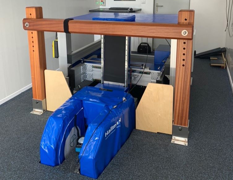
- Hallmarq Racing WA Standing Equine MRI.
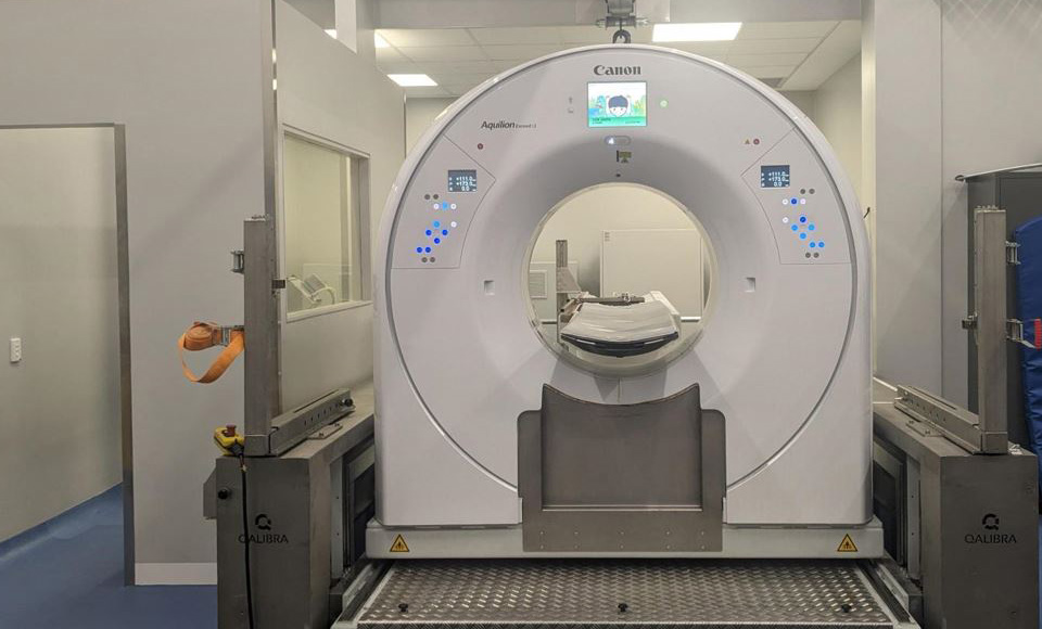
- Qalibra Canon Exceed 160 slice Large Bore (90cm) Standing Equine CT.
These imaging modalities are provided either as referral-only, direct-accession outpatient services, or for TAHMU patients, with the aim to provide these services to as wide a population of horses, horse owners and referring veterinarians as possible. Horses have brief physical examinations prior to sedation. Images are acquired by TAHMU staff and the horses discharged back into your care on the same day (barring emergencies).
All reporting is provided by specialist radiologists, and further clinical opinions can be provided as necessary. Alternatively, horses can be referred to TAHMU for further evaluation and treatment.
MRI (Magnetic Resonance Imaging)

CT (Computed Tomography)
CT imaging is useful for investigating the head and neck, including dental abnormalities, traumatic injuries of the skull, nasal passage problems, headshaking, and neurological diseases. CT images are also excellent for assessing bone injuries of the limbs, such as complex fractures and stress fractures which aren’t visible on radiographs.
The resulting images are extremely helpful for our surgeons to assist with planning of orthopaedic cases requiring surgical treatment. The Animal Hospital at Murdoch University is lucky to have the most advanced standing equine CT system in the world.


-sitting-in-front-of-tah-signage.tmb-300-sqcrop.jpg?Culture=en&sfvrsn=eb6587e5_1)